


Retrocalcaneal Bursitis Treatment & Doctors in Bangalore
- Home
- Conditions
- Foot & Ankle Pain
- Retrocalcaneal Bursitis
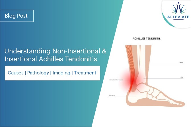
The most likely bursa around the heel that is responsible for heel pain is the retrocalcaneal bursa. This pain results when this bursa( sac like structure lined by a thin layer of synovial fluid, functions to reduce friction between surfaces) gets filled up with inflammatory fluid and results in Retrocalcaneal Bursitis. The location of the bursa is between the calcaneum and the anterior surface of the Achilles tendon. There are two bursae in this area. Deep to the Achilles tendon lies the sub tendinous or retrocalcaneal bursa. Superficial to the Achilles lies the subcutaneous calcaneal bursa (This bursa lies between posterior surface of the Achilles tendon and the skin). Inflammation of either of these bursae can lead to pain at the posterior heel and ankle region.
Treatment for Retrocalcaneal Bursitis
Relevant Clinical Anatomy & Pathogenesis
The retrocalcaneal bursa is housed over the posterio-superior prominence of the heel bone right under the Achilles tendon and it’s lateral expansions. The calcaneum, fibrocartilaginous walls of the retrocalcaneal burial and the insertion of the Achilles tendon form an ‘Enthesis Organ’. Conceptually, at the site of the Achilles tendon insertion, the bursae and the bone are so intimately linked that a small prominence of the calcaneum will greatly increase the chances of mechanical irritation of the bursa.
Etiology
- Ill fitting footwear and especially switching to flat shoes after a prolonged period of using high heels can predispose individuals to Retrocalcaneal bursitis.
- Repetitive micro trauma to the bursa due to excessive loading as is seen athletes who tend to overstrain can be a predisposing factor.
- Haglund deformity
- Alteration of the joint axis
Sign & Symptoms
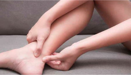
- Troubling pain at the back of the hill especially while walking or running uphill.
- Standing up on toes can aggravate the pain.
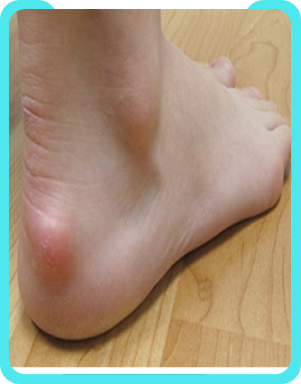
- Tenderness on palpation at the back of heel.
- Swelling often associated with warmth and redness at the back of heel
- Aggravation of pain with calf loading activities.
Clinical Assessment & Investigations
- Careful history and a thorough examination to look for local swelling, tenderness, evaluation of the tendon, detecting any bony prominence as well as locating the area of maximum tenderness with palpation.
- Tightening of proximal soft tissue, stiffness in joint and any biomechanical abnormalities Should be looked for as these can make one anatomically predisposed to retrocalcaneal bursitis.
- X rays may show the presence of a bony prominence in the postern-superior aspect of the calcaneum. This is termed as the Haglund deformity.
- The retrocalcaneal recess often looks normal on weight bearing lateral x rays making their use limited in aiding the diagnosis.
- Patients may show the absence of normal radiolucency at the posteroinferior corner of the Kager fat pad, (blunting). There may be associated calcaneal erosion seen.
- Musculoskeletal Ultrasound is a great tool in imaging the bursa, appreciating any bursal thickening, inflammation, health of the adjoining soft tissues and is also mighty helpful in carrying out Interventional Pain Management and Regenerative Procedures for the same.
- Magnetic resonance imaging (MRI) can demonstrate bursal inflammation but does not o er much greater information helping in the diagnosis.
- Gout, rheumatoid arthritis and seronegative spondyloarthropathies
Differential Diagnosis
- Haglund Deformity
- Achilles Tendonitis
- Partial rupture of the Achilles tendon
- Plantar Fasciitis
- Posterior Ankle Impingement
Treatment
Physical Therapy :
- Icing of the heel and posterior part of ankle can be performed several times a day during active inflammation for stretches of 15-20 minutes.
- Exercises involving the stretching of the Achilles tendon are advised. These can help in relieving the impingement on the subten dinous bursa.
- Calf stretch keeping the back leg straight with the heel placed on the ground one should lean forward against a wall. The front leg has a bend in the knee . To stretch the gastrocnemius complex (calf muscles) and the Achilles tendon, hips have to be pushed towards the wall in a controlled manner. Position is to be held for 10 seconds before relaxing. Exercise should be repeated around 20 times for each foot and this should give the adequate stretch in the calf muscles.
Corticosteroid Injection : Traditionally a lot of people have received blind corticosteroid injections by clinicians for this condition. In our practice, we advocate precise ultrasound guided injections for really painful bursitis. This makes sure that the site of the injection is the bursa avoiding steroid infiltration in nearby tissues.
Platelet Rich Plasma & Prolotherapy : TFor most cases of retrocalcaneal bursitis encountered at ALLEVIATE, we follow a Comprehensive Platelet Rich Plasma and Prolotherapy treatment for the same. Patient usually respond well to treatment in two to three sessions of the same. Though our treatment is coupled with a rigorous multidisciplinary approach focussing on exercise, physical therapy, nutrition and weight management as well. SURGERY- For extremely chronic and resistant cases a Bursectomy might be undertaken.( Rarely done)
FAQs - Retrocalcaneal Bursitis
Retrocalcaneal bursitis is an inflammation of a bursa between the Achilles tendon and heel bone. This inflammation can occur by friction or shearing of the bursa, which can be caused by shoes that squeeze your heel or cause you to change your gait.
There are many possible causes of Retrocalcaneal Bursitis.
It is more common in people who do a lot of running or other high-impact activities. In some cases, it may be due to pre-existing injury or condition.
In other cases, it is caused by an injury to the heel. The injury might be because of falling, especially if the foot is twisted or if a person is wearing high heels or platform shoes. Ankle sprains, particularly in dancers, can also cause Retrocalcaneal.
The symptoms of Retrocalcaneal Bursitis include:
- Pain in the heel
- Pain in the ankle
- Inflammation around the heel
- Redness or swelling of the heel
- Heel tenderness
- Pain when the ankle is bent or pushed back
- Aggravate pain while standing up on toes
The diagnosis of retrocalcaneal bursitis is done by careful clinical assessment and investigations, including a manual exam and possibly a test to detect swelling, tenderness, tendon, any bony prominence and locating the area of maximum tenderness with palpation.
The diagnostic tests for retrocalcaneal bursitis includes X-ray, CT scan, bone scan or MRI.
Treatment options for retrocalcaneal bursitis are as follows:
- Physical Therapy – Icing, Exercise, Calf Stretch
- Corticosteroid Injection
- Platelet Rich Plasma & Prolotherapy
- Surgery
Physical therapy for retrocalcaneal bursitis involves the use of non-surgical physical therapy intervention to reduce discomfort and decrease the risk of chronic or recurring pain between the heel bone and the Achilles tendon.
Stretching and strengthening the calf muscles is a mainstay of treating bursitis. Calf stretching exercises are performed to relieve pain and inflammation in the heel and arch of the foot.
Injections of corticosteroids (steroids) into the retrocalcaneal bursa (back of the heel) can provide pain relief for this condition.
Platelet Rich Plasma (PRP) & Physiotherapy has shown to be effective at treating retrocalcaneal bursitis. With this treatment, most patients have successfully responded to treatment. The treatment can be done in two to three sessions.
Electrical modalities have been tried but are not hugely recommended by doctors.
Retrocalcaneal Bursitis can mostly be treated with non-surgical intervention, which has a high success rate at Alleviate Pain Clinic. For extreme chronic and resistant cases, surgery is rarely done.
Video Spotlight
Blog
Surgery-Free Solutions
Expert Tips for Pain Management

Testimonials
Words From Our Patients
The treatment was very good and the doctor Faraz Ahmed was very kind to the patient and explained clearly the procedure of knee bilateral ha & treatment And we were advised to do physiotherapy. We are very much satisfied. We would recommend this alleviate pain clinic. Thank you
Got treatment of Treatment and HA for right knee arthritis a month ago and finding good relief from pain. Was treated by Dr Swagtesh Bastia who explained very well about the injections and the treatment was painless. The front desk staff were very kind and very helpful and physiotherapy was also done expertly, overall good experience
Alleviate Pain Management clinic has been a godsend for my mom's knee pain. She has been treated by Dr. Wiquar Ahmed. The attentive staff provided personalised care, and after her treatment, she's feeling remarkably better. Thank you for giving my mom the relief she deserves!
The clinic is super clean with a great OT and most importantly all the staff here are very helpful and considerate. My gratitude to Dr Roshan, the nurses, and support staff - they were always available to assist with any issues post procedure and they even made an extra effort to make a home visit for a follow up check-up. This team here is the perfect example of healing and care with a human touch. Thank you!!!!!
My wife had knee pain I have visited alleviate pain and consulted doc santhoshi now she is able to walk pain free and can do her daily activity than before.the physiotherapist here Dr akhila also helped her with few exercises and the staff here Abdul explained all the procedures well . Thank you PPL can visit here for pain relief
