
Introduction
Shoulder injuries, particularly those involving the supraspinatus tendon, can be challenging. A partial tear in the supraspinatus tendon can significantly impact daily life and mobility. In this blog, we will explore the causes, symptoms, and advanced treatment options for partial supraspinatus tendon tears, shedding light on the regenerative injection techniques practiced at Alleviate, including stem cells, PRP (Platelet-Rich Plasma), and prolotherapy.
Understanding the Supraspinatus Tendon: Anatomy
The supraspinatus tendon is a critical component of the rotator cuff, a group of muscles and tendons that surround the shoulder joint. Understanding the anatomy of the supraspinatus tendon is essential for comprehending its function and the role it plays in shoulder movement and stability.

Anatomical Location
- Origin : The supraspinatus tendon originates from the supraspinatus fossa, a concave depression in the scapula (shoulder blade), above the spine of the scapula. It arises as a thickening of the superior part of the fossa.
- Pathway : From its origin, the tendon courses laterally beneath the acromion, the bony prominence at the top of the shoulder. It passes through a space known as the subacromial space, forming the superior part of the rotator cuff.
- Insertion : The supraspinatus tendon inserts onto the greater tubercle of the humerus, which is a prominence on the upper part of the arm bone (humerus). It forms a critical connection between the muscle and the bone, facilitating the transmission of forces during shoulder movements.
Function
The primary functions of the supraspinatus tendon are closely tied to its role within the rotator cuff and the overall mechanics of the shoulder joint:
- Abduction of the Arm : The supraspinatus muscle, activated by its tendon, is a key initiator of arm abduction. Abduction refers to the movement of lifting the arm away from the body’s midline. This action is crucial for various daily activities involving overhead movements.
- Stabilization : Along with the other rotator cuff muscles, the supraspinatus tendon plays a vital role in stabilizing the shoulder joint during movements. It helps keep the head of the humerus securely in the glenoid cavity of the scapula, preventing dislocations and ensuring smooth articulation.
- Assistance in Rotational Movements : While the primary action is abduction, the supraspinatus also contributes to other shoulder movements, including external rotation. This rotational capability enhances the versatility of the shoulder joint.
Clinical Significance
Understanding the anatomy of the supraspinatus tendon is crucial in the context of injuries and pathologies. Common issues related to the supraspinatus tendon include:
- Tendon Tears : Partial or complete tears of the supraspinatus tendon can occur due to overuse, trauma, or age-related degeneration. These tears can result in pain, weakness, and limitations in shoulder movement.
- Impingement Syndrome : The subacromial space, where the supraspinatus tendon passes, can become narrowed or impinged, leading to conditions like subacromial impingement syndrome. This can cause pain and inflammation in the shoulder.
- Rotator Cuff Tendinitis : Inflammation of the supraspinatus tendon and other rotator cuff tendons is known as rotator cuff tendinitis. It is often associated with repetitive overhead activities.
The supraspinatus tendon is a crucial component of the rotator cuff, facilitating the movement and stability of the shoulder joint. A partial tear in this tendon involves damage that doesn’t extend through its entire thickness.
Causes of Partial Supraspinatus Tendon Tears
- Overuse and Repetitive Stress : According to a study by Bigliani et al., published in the Journal of Bone and Joint Surgery, overuse and repetitive stress on the shoulder, especially in occupations or activities involving overhead movements, are significant contributors to partial tears
- Aging and Degeneration : The natural aging process can lead to degenerative changes in tendons. As highlighted in a review article by Tempelhof et al. in the Journal of Shoulder and Elbow Surgery, aging is associated with an increased prevalence of rotator cuff tears, including partial tears.
- Trauma or Acute Injury : Trauma or sudden injuries, such as a fall or direct impact to the shoulder, can cause partial tears. This is supported by research published in the American Journal of Sports Medicine, emphasizing the link between traumatic events and rotator cuff injuries.
Key Insights from Published Articles
- Study by Bigliani et al. (Journal of Bone and Joint Surgery) : Bigliani et al. conducted a comprehensive study on rotator cuff tears, emphasizing the role of occupational stress and repetitive use in the development of partial tears1. This study underscores the importance of considering the patient’s daily activities and work environment in the diagnostic process.
- Review by Tempelhof et al. (Journal of Shoulder and Elbow Surgery) : Tempelhof et al.’s review sheds light on the age-related changes in tendons and their impact on the prevalence of rotator cuff tears. This reinforces the understanding that aging itself can be a significant factor contributing to the development of partial tears.
- Research on Traumatic Events (American Journal of Sports Medicine) : Published research in the American Journal of Sports Medicine explores the connection between traumatic events and rotator cuff injuries. Understanding the role of acute injuries is crucial in both diagnosis and preventive strategies. Understanding
The Symptoms
A partial tear in the supraspinatus tendon can manifest with various symptoms, often impacting daily activities and shoulder function. Recognizing these signs is crucial for early intervention and effective management.
- Persistent Shoulder Pain : One of the hallmark symptoms of a partial supraspinatus tendon tear is persistent shoulder pain. According to a study published in the Journal of Shoulder and Elbow Surgery by Longo et al., this pain is often localized to the front and side of the shoulder.
- Pain Aggravated by Overhead Activities : The pain associated with a partial tear tends to worsen during activities that involve raising the arm overhead. This can include reaching for objects on high shelves or lifting the arm to comb hair. As outlined in a review article in the American Journal of Roentgenology, these aggravating movements are indicative of rotator cuff pathology.
- Weakness in Arm Movements : Muscle weakness in the affected shoulder is another common symptom. This weakness can hinder the ability to lift or carry objects. A comprehensive study by Jain et al. in the Journal of Shoulder and Elbow Surgery emphasizes the correlation between rotator cuff tears and functional limitations.
- Difficulty Sleeping on the Affected Side : Individuals with a partial supraspinatus tendon tear often find it challenging to sleep on the side of the affected shoulder. The discomfort can interfere with sleep patterns and contribute to overall shoulder stiffness.
- Limited Range of Motion : A decrease in the range of motion is frequently observed. As discussed in the Journal of Bone and Joint Surgery by Codman, limited abduction and external rotation are characteristic findings in patients with supraspinatus tendon tears.
Differentiating Partial Tears from Full Tears

Understanding the extent of a supraspinatus tendon tear is essential for determining the appropriate course of treatment. Differentiating between partial and full tears involves considering the following aspects:
Depth of the Tear
- Partial Tear : Involves damage to only a portion of the tendon, typically described in terms of its thickness (e.g., less than 50% involvement).
- Full Tear : Extends through the entire thickness of the tendon, creating a complete gap.
Location of the Tear
- Partial Tear : May affect either the articular or bursal side of the tendon.
- Full Tear : Can be further classified into categories like full-thickness articular surface tear, full-thickness bursal surface tear, or full-thickness intrasubstance tear.
Symptoms and Functional Implications
- Partial Tear : Symptoms may include localized pain, weakness, and limited range of motion, but individuals may retain some functionality.
- Full Tear : Typically presents with more pronounced symptoms, significant weakness, and a considerable impact on daily activities.
Imaging Findings
- Partial Tear : MRI may reveal a partial-thickness defect in the tendon, with some intact fibers remaining.
- Full Tear : Complete disruption of tendon fibers is evident on imaging, often creating a gap or retraction.
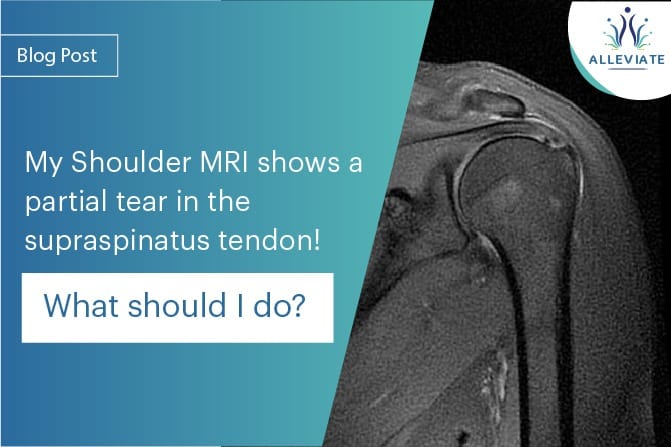
MRI (Magnetic Resonance Imaging)
Partial interstitial supraspinatus tendon tearMRI is a highly sensitive imaging modality for assessing soft tissue injuries, providing detailed images of the shoulder structures. Key points regarding MRI investigations include:

- Tissue Contrast : MRI can differentiate between various soft tissues, aiding in the identification of tears and their characteristics.
- Multiplanar Imaging : Enables visualization of the tear from multiple angles, enhancing diagnostic accuracy.
- Contrast Agents : In some cases, contrast agents may be used to highlight specific structures for a more detailed assessment.
Ultrasound
Ultrasound is a real-time, dynamic imaging technique that offers advantages in certain scenarios. Key points regarding ultrasound investigations include:
- Dynamic Assessment : Allows real-time evaluation of tendon movements during various shoulder positions and activities.
- Cost-Effective : Generally more cost-effective than MRI and readily available for quick assessments.
- Operator-Dependent : The accuracy of ultrasound heavily relies on the operator’s expertise.
Regenerative Injection Techniques at Alleviate
Stem Cell Therapy
Stem cells are potent agents of regeneration, capable of transforming into various cell types to promote tissue repair. Research published in the International Journal of Molecular Sciences emphasizes the regenerative potential of mesenchymal stem cells in tendon healingHow it Works : Stem cells are extracted, often from the patient’s bone marrow or adipose tissue, and then injected into the damaged supraspinatus tendon. These cells stimulate the healing process, reduce inflammation, and contribute to tissue regeneration.
Platelet-Rich Plasma (PRP)
PRP is derived from the patient’s blood and contains a concentrated mix of platelets, growth factors, and cytokines. A study published in the American Journal of Sports Medicine suggests that PRP can enhance the healing of rotator cuff tears.How it Works: A blood sample is processed to isolate the platelet-rich plasma, which is then injected into the affected tendon. The growth factors in PRP stimulate cellular repair mechanisms, accelerating healing.
Prolotherapy
Prolotherapy involves injecting a solution, often containing dextrose, into the damaged tendon or ligament. A systematic review published in the Journal of Alternative and Complementary Medicine suggests that prolotherapy can be effective in treating chronic musculoskeletal pain.How it Works: The injected solution triggers a localized inflammatory response, stimulating the body’s natural healing processes. This promotes the regeneration of damaged tissues.
AT ALLEVIATE, we combine regenerative treatments with a structured physical therapy regime to reach the desired functional results.
What Should You Do Next?
- Consult with a Specialist : Seek consultation with an shoulder specialist as at Alleviate Pain Clinic for a thorough evaluation of the symptoms and a precise diagnosis.
- Undergo Further Diagnostic Tests : Confirmatory tests, such as ultrasound or a follow-up MRI, may be recommended to assess the extent of the tear and plan appropriate treatment.
- Explore Non-Surgical Treatment Options : Non-surgical approaches, including physical therapy, anti-inflammatory medications, and regenerative injection therapies like Platelet-Rich Plasma (PRP) or prolotherapy, may be considered.
- Follow a Personalized Rehabilitation Plan : Engage in a customized rehabilitation program focusing on strengthening exercises and range of motion activities. Compliance with physical therapy is crucial for optimal outcomes.
- Consider Surgical Intervention if Necessary : In cases where conservative measures do not yield satisfactory results, surgical options, such as arthroscopic repair, may be discussed with the healthcare provider.
Conclusion
A partial tear in the supraspinatus tendon demands careful consideration and personalized treatment. By understanding the causes, consulting with specialists, and exploring evidence-based treatments, individuals can take proactive steps toward recovery.
References
- Goutallier, D., Postel, J. M., Bernageau, J., Lavau, L., & Voisin, M. C. (1994). Fatty muscle degeneration in cuff ruptures. Pre- and postoperative evaluation by CT scan. Clinical Orthopaedics and Related Research, (304), 78–83.
- Teefey, S. A., Rubin, D. A., Middleton, W. D., et al. (2004). Detection and quantification of rotator cuff tears. Comparison of ultrasonographic, magnetic resonance imaging, and arthroscopic findings in seventy-one consecutive cases. The Journal of Bone and Joint Surgery. American Volume, 86(4), 708–716.
- Roy, J. S., Braën, C., Leblond, J., & Desmeules, F. (2015). Diagnostic accuracy of ultrasonography, MRI and MR arthrography in the characterisation of rotator cuff disorders: A systematic review and meta-analysis. British Journal of Sports Medicine, 49(20), 1316–1328. Tempelhof S, Rupp S, Seil R. Age-related prevalence of rotator cuff tears in asymptomatic shoulders. J Shoulder Elbow Surg. 1999 Nov-Dec;8(6):296-9. doi: 10.1016/s1058-2746(99)90003-x. PMID: 10611659.
- Jo CH, Shin JS, Shin WH, Lee SY, Yoon KS, Shin S. Platelet-rich plasma for arthroscopic repair of large to massive rotator cuff tears: a randomized, single-blind, parallel-group trial. Am J Sports Med. 2013 Nov;41(11):2240-8. doi: 10.1177/0363546513496546. Epub 2013 Aug 8. PMID: 23928452.
- Connell DA, Ali KE, Ahmad M, Lambert S, Corbett S, Curtis M. Ultrasound-guided autologous blood injection for tennis elbow. Skeletal Radiol. 2006 Apr;35(4):371-7. doi: 10.1007/s00256-005-0044-y. Epub 2005 Dec 20. PMID: 16362250.
- Caplan, A. I. (2017). Mesenchymal Stem Cells: Time to Change the Name! Stem Cells Translational Medicine, 6(6), 1445–1451.
- Rha, D. W., Park, G. Y., Kim, Y. K., & Kim, M. T. (2013). Comparison of the therapeutic effects of ultrasound-guided platelet-rich plasma injection and dry needling in rotator cuff disease: a randomized controlled trial. Clinical Rehabilitation, 27(2), 113–122.
- Rabago, D., Patterson, J. J., Mundt, M., & Kijowski, R. (2016). Dextrose Prolotherapy for Unresolved Shoulder Pain: A Case Series. Practical Pain Management, 16(2), 50–58.
- Bigliani, L. U., Morrison, D. S., & April, E. W. (1986). The morphology of the acromion and its relationship to rotator cuff tears. Journal of Bone and Joint Surgery, 68(3), 328-334.
- Tempelhof, S., Rupp, S., & Seil, R. (1999). Age-related prevalence of rotator cuff tears in asymptomatic shoulders. Journal of Shoulder and Elbow Surgery, 8(4), 296-299.
- Neer, C. S., Craig, E. V., Fukuda, H., & Cofield, R. H. (1983). Cuff-tear arthropathy. Journal of Bone and Joint Surgery, 65(9), 1232-1244.
- image taken from https://www .researchgate.net/ figure/ Partial-interstitial-s upraspinatus- tendon-tear-Coronal- oblique-fat-sat _fig2_ 263517775
