


Plantar Fasciitis Introduction
Plantar fasciitis is the most common cause of heel pain in adult population requiring medical attention. Statistics show that there is a good chance that 1 in every 10 people will experience the symptoms of plantar fasciitis at some point in their life.[5] Historically plantar fasciitis has had quite a few synonyms namely chronic plantar heel pain, heel spur syndrome, runner’s heel, painful heel syndrome and calcaneal periostitis.
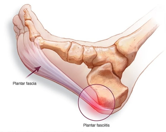
Clinical Anatomy With Pathological Features
A Fibro-fatty fat pad sits in between the heel bone and the plantar skin. The posterior tuberosity of the calcaneum gives out a medial and lateral process. The posterior tuberosity of calcaneum has a medial and a lateral processes. The medial process gives attachment to the Flexor digitorum brevis (FDB), Abductor hallucis (AH), and the medial head of Quadratus plante (QP) as well as the central band of plantar fascia.
The plantar fascia or deep fascia of the sole, proximally has a direct fibrocartilaginous attachment to the calcaneum (an enthesis), whose central band is constant along with medial and lateral band. It has a triangular shape and develops from the medial process of the calcaneal tuberosity, and diverges distally at mid-metatarsal level into five separate strands, which are attached at the forefoot onto the plantar skin, the base of proximal phalanges (via plantar plate), the metatarsophalangeal(MTP) joints via the collateral ligaments and deep transverse metatarsal ligaments.[6]
Heel skin is innervated by the medial calcaneal nerve which may present with heel pain if compressed proximally (such as in tarsal tunnel syndrome). The histological features of plantar fasciitis are more suggestive of degeneration rather than inflammation. Histology in many cases is consistent with Fascitis and Fasciosis. .information about its pathogenesis is still limited, and its histological changes are suggestive of degeneration rather than inflammation. The fascia is usually markedly thickened and gritty.[8]
There is evidence of spur formation in the loose connective tissue, spur trabecular forming perpendicular to the long axis and fibrocartilage orientation away from the lines of traction.
FUNCTIONS OF THE PLANTAR APONEUROSES/FASCIA
The aponeurosis is important for
- Guarding deeper structures of the foot, such as nerves and vessels.
- Maintenance of the longitudinal arch of the foot.
- Provides attachment for muscles.
- Prevents excessive dorsiflexion.
- Distributes plantar pressure during static and dynamic loading.
Risk Factors associated with PF
Knowledge of the risk factors of plantar fasciitis can help plan preventative and treatment strategies.
- Obesity finds it’s association with at least 60-70% of the patients suffering with plantar fasciitis. Research shows a high correlation between increased body weight and plantar fasciitis whereas the correlation of PF( Plantar Fasciitis) with height has not shown much statistical significance.
- Calcaneal spurs are commonly associated with PF.
- Prolonged standing has been considered by some as being a contributing factor.
- Kiber et al attributed deficits in flexibility of the plantar flexor muscles leading to greater fascia stretching.[9]
- Cheung et al propose that intense muscular contractions of the plantar flexor muscles can lead to indirect stretching of the fascia predisposing one to PF.[10]
- Pronated foot posture and over pronation of the foot are also believed to be possible risk factors.[11]
Clinical Examination
The diagnosis of plantar fasciitis is essentially clinical.[12,13] but Ultrasound imaging can give objective information on the health of the fascia. Tenderness is usually found on the medial side of the heel. Patient classically describes more pain after a period of inactivity like sleep. Symptoms tend to get better during the day with activity again tending to worsen at night. Prolonged and increased weight bearing can be aggravating factors. Around 80% of the patients have associated tightness in the Achilles tendon. In some cases the pain may radiate down to the foot.
Imaging studies
- Lateral X rays of the of the ankle is a good modality for assessment of heel spur, thickness of plantar fascia, and the quality of fat pad.
- Ultrasound examination in experienced hands can be very helpful when the diagnosis is unclear.[14,15] In the literature, normal thickness of the plantar fascia when measured in ultrasound varies in range (mean 2–3 mm). People with chronic heel pain are likely to have a thickened plantar fascia with associated fluid collection, and that thickness values >4.0 mm are diagnostic of plantar fasciitis.[6]
- Plantar fascia thickness values have also been used to gauge the the efficacy of treatments and a significant correlation has been found between decreased plantar fascia thickness and improvement in symptoms.[16,17,18]
- MRI can be helpful in resistant or doubtful cases as it can throw light on other conditions like as tarsal tunnel syndrome, soft tissue and bone tumors, osteomyelitis, subtalar arthritis, and stress fracture.[17,18]
Differential diagnosis
- Plantar Fascia Rupture
- Fat Pad syndrome
- Calcaneal stress fracture
- Calcaneal Bursitis/ Policeman’s heel
- Boxter’s nerve entrapment
- Medial Calcaneal Nerve Compression
- Seronegative Arthropathies
- Spinal Stenosis and L5-S1 Nerve Root Irritation
- Tumor
Treatment
The natural course of plantar fasciitis may last from 8-18 months making it a considerable duration of daily discomfort for the patient. Hence timely intervention can help significantly shorten the period of discomfort.
NSAIDs like naproxen or etodolac in the the acute stage are helpful in getting the pain and inflammation down.
Stretching
Calf stretching has been advocated by many experts as an important excercise in rehabilitation. Stretching may be done in the calf or plantar region. These stretches are recommended two to three times a day.
Night splints
Night splinting is done to encourage a neutral position of the ankle at night allowing passive stretching of the plantar fascia during sleep. They might be advised for 1-3 months depending on the severity.
Orthosis
Orthoses were traditionally used to reduce abnormal foot pronation. Prefabricated and customised insoles work by supporting the longitudinal arch thus helping relieving the pain.
Extra-corporeal shock wave therapy
Extra-corporeal shock wave therapy (ESWT) is believed to bring about deep tissue cavitation effect causing micro rupture of capillaries, leakage of chemical mediators, and promotion of neovascularization of the damaged tissue.[6] ESWT is carried out under intravenous sedation with or without local anaesthetic infiltration..[19,20] This modality is indicated if there is failure of other conservative measures.
Corticosteroid injection
Resistant cases are often given blind or image guided corticosteroid injections due to it’s potent anti- inflammatory effect. Ultrasound guided injections are much safer and precise [21] guiding the clinician deep into the fascia thus almost eliminating the risk of fat pad necrosis or fascial rupture. Medial approach is usually preferred as it is less painful.
AT ALLEVIATE we are primarily focussed on treating the chronic cases of plantar fasciitis we encounter with, Autologous platelet rich plasma (PRP) with Comprehensive prolotherapy.
Addressing the thickened fascia with Platelet Rich Plasma, the adjoining Achilles tendon and ankle ligaments are subjected to comprehensive prolotherapy. Ideally we try to do around three sessions of this treatment at intervals of 2-3 weeks.
We have found incredible results in a vast majority of patients who underwent the complete treatment. They reported greatly reduced pain and much improved function. Moreover as opposed to steroids, we have found the positive effects of regenerative treatment to be much more lasting . Our treatments are always coupled with a multidisciplinary approach.
Surgery
Recalcitrant cases where symptoms persist for more than 6–12 months, even after adequate conservative treatment are usually selected for Open or endoscopic plantar fascia release. Nerve conduction studies to rule out posterior tibial nerve compression should be done preoperatively.
