

Introduction
Osteoarthritis of the knee is a prevalent joint disorder that affects millions of people worldwide. This degenerative condition leads to progressive cartilage loss in the knee joint, causing pain, stiffness, and reduced mobility. In this blog, we will delve into the etiopathogenesis, epidemiology, radiological features, and pain generators associated with osteoarthritis of the knee, shedding light on its impact and management.
What is Osteoarthritis of the Knee?
Osteoarthritis is a chronic joint condition characterized by the gradual degradation of articular cartilage, the protective tissue that covers the ends of bones within a joint. In the case of knee osteoarthritis, the cartilage in the knee joint undergoes wear and tear, leading to the exposure of underlying bones. This results in inflammation, pain, and other symptoms that significantly impact a person’s quality of life.
Etiopathogenesis of Knee Osteoarthritis
The exact cause of knee osteoarthritis is not entirely understood. It is considered a multifactorial condition with several contributing factors, including:
- Age : The risk of developing knee osteoarthritis increases with age, as cartilage repair mechanisms become less efficient over time.
- Hereditary : Certain genetic factors may make some individuals more susceptible to developing knee osteoarthritis.
- Previous Trauma : Previous injuries to the knee, such as ligament tears or fractures, can predispose individuals to osteoarthritis.
- Obesity : Excess body weight places added stress on the knee joints, accelerating cartilage deterioration.
- Gender : Women are more prone to knee osteoarthritis than men, especially after menopause.(1)
- Joint Malalignment : Poor joint alignment or abnormal joint mechanics may contribute to uneven cartilage wear. (2)
- Chronic Inflammation : Chronic low-grade inflammation in the joint can contribute to cartilage breakdown.
- Laxity of knee ligaments– Varus or Valgus laxity can be a predisposing factor in many individuals.
Epidemiology of Knee Osteoarthritis
Knee osteoarthritis is a prevalent condition, and its prevalence increases with age. Some key epidemiological facts include:
- Global Impact: Knee osteoarthritis is one of the most common musculoskeletal disorders worldwide, affecting millions of individuals.
- Age-Related: The prevalence of knee osteoarthritis rises significantly with age, especially in individuals over 60 years old. The age standardized prevalence of radiographic knee OA in adults age ≥ 45 was 19.2% among the participants in the Framingham Study and 27.8% in the Johnston County Osteoarthritis Project (3). In the third National Health and Nutrition Examination Survey (NHANES III), approximately 37% of participants age >60 years or older had radiographic knee OA (3).
- Gender Differences: Women are more likely to develop knee osteoarthritis than men, especially after the age of 50.
- Obesity and Sedentary Lifestyle: Obesity and a sedentary lifestyle are significant risk factors for knee osteoarthritis.
Radiological Features of Knee Osteoarthritis
Radiological imaging plays a crucial role in diagnosing and assessing knee osteoarthritis. Common radiological features include:
- Joint Space Narrowing : X-rays reveal a decrease in the joint space between bones due to cartilage loss.
- Osteophytes : Bone spurs may form at the joint margins in response to the cartilage breakdown.
- Subchondral Sclerosis : Increased bone density beneath the affected cartilage is a characteristic feature.
- Cysts : Fluid-filled cysts may develop within the bone near the joint space.
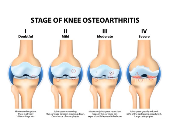
KELLGREN LAWRENCE GRADING OF KNEE OSTEOARTHRITIS
AP radiographs of the knee presented in the original Kellgren-Lawrence article [4]. (A) Representative knee radiograph of KL classification Grade 1, which demonstrates doubtful narrowing of the joint space with possible osteophyte formation. (B) Representative knee radiograph of KL classification Grade 2, which demonstrates possible narrowing of the joint space with definite osteophyte formation. (C) Representative knee radiograph of KL classification Grade 3, which demonstrates definite narrowing of joint space, moderate osteophyte formation, some sclerosis, and possible deformity of bony ends. (D) Representative knee radiograph of KL classification Grade 4, which demonstrates large osteophyte formation, severe narrowing of the joint space with marked sclerosis, and definite deformity of bone ends.
Pain Generators in Knee Osteoarthritis
Knee pain in osteoarthritis can arise from various sources :
- Inflamed Synovium: The synovial membrane lining the joint can become inflamed, causing pain.
- Cartilage Damage: As cartilage wears away, the underlying bone is exposed, leading to pain and inflammation.
- Bone Spurs: Osteophytes or bone spurs can irritate surrounding tissues, contributing to pain.
- Ligament and Tendon Strain: Strain on ligaments and tendons around the knee joint can result in pain.
Conclusion
Osteoarthritis of the knee is a common and complex joint disorder with various contributing factors. It predominantly affects the aging population but can also impact younger individuals due to various risk factors. Radiological imaging is crucial in diagnosing knee osteoarthritis, and pain can originate from different structures within the joint. Early diagnosis, lifestyle modifications, and appropriate management are essential in alleviating symptoms and improving the quality of life for those living with knee osteoarthritis.
If you are suffering with knee pain due to osteoarthritis, we urge you to visit us at ALLEVIATE PAIN CLINIC to get a detailed understanding of your condition and the treatment protocol that would be suited for your stage of the the disease.
References
- Srikanth VK, Fryer JL, Zhai G, Winzenberg TM, Hosmer D, Jones G. A meta-analysis of sex differences prevalence, incidence and severity of osteoarthritis. Osteoarthritis Cartilage. 2005;13(9):769–81. [PubMed] [Google Scholar]
- Sharma L, Song J, Felson DT, Cahue S, Shamiyeh E, Dunlop DD. The role of knee alignment in disease progression and functional decline in knee osteoarthritis. Jama. 2001;286(2):188–95. [PubMed] [Google Scholar]
- Lawrence RC, Felson DT, Helmick CG, et al. Estimates of the prevalence of arthritis and other rheumatic conditions in the United States. Part II. Arthritis Rheum. 2008;58(1):26–35. [PMC free article] [PubMed] [Google Scholar]
- Kellgren JH, Lawrence JS. Radiological assessment of osteo-arthrosis. Ann Rheum Dis. 1957;16:494–502. doi: 10.1136/ard.16.4.494. [PMC free article] [PubMed] [CrossRef] [Google Scholar]
