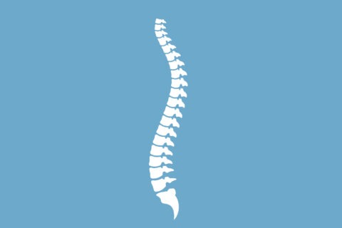
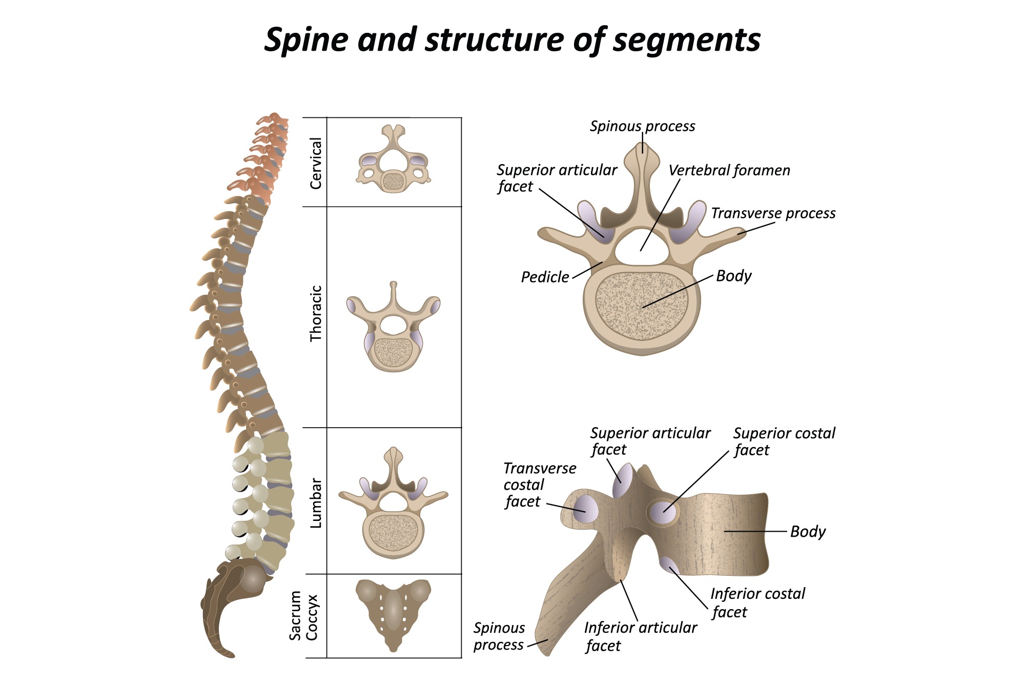
The Spinal Column is also called the vertebral column. The bones in the spine are called vertebrae (ver-ta-bray). The column starts at the base of the skull and continues to the pelvis. Alternate layers of bone (vertebrae) and cartilage (car-til-ledge, the intervertebral discs) stack vertically one on top of the other in the spinal column. The lattice-like structure of the cancellous bone (cancel-lus, the spongy interior) in a vertebra absorbs external pressure. The human spine has natural curvatures. When you look at a back from behind, the spine should be straight and centered over the pelvis. However, when you look at the spine from the side, the curves are designed to maintain balance as the spine is behind organs in the chest and abdomen. The spine has two alternating curves to create an “S” like shape. In the neck and low back there is normally an inward curvature or sway back known as lordosis. In the thoracic spine and sacrum there is an outward curvature known has kyphosis or hunchback. These curves normally balance out each other so that when the patient stands they are well balanced with their head straight above their hips when viewed from the side. Standing in this position minimizes the effect of gravity and allows the patient to stand with the best posture and use the least energy when moving or walking. There are seven cervical (C) vertebrae, twelve thoracic (T) vertebrae, and typically five lumbar (L) vertebrae.
Anatomy of the Spine
Anatomy of the Spine
Anatomy of the Spine

-
VERTEBRAE
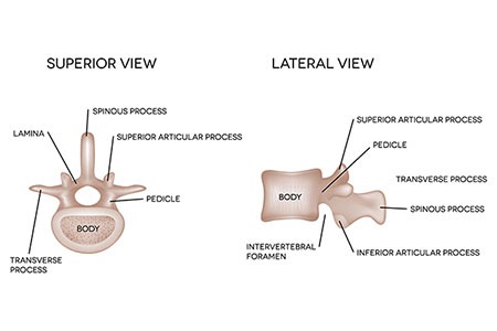 The spine is made of a column of bones. A round bone called a vertebral body forms each bone. The body is located in the front of the spine. A bony ring attaches to the back of the vertebral body, forming a canal for the spinal cord and nerves. This bony ring is formed by two sets of bones. One set called the pedicle bone attaches to the back of each vertebral body to the side, transverse processes. Processes are outgrowths of bone and then subsequently named for where the outgrowth occurs. A lamina (Latin for plate) bone connects to the other end of the pedicle, one on the left and one on the right to connect the transverse process with the spinous process. The spinous process is your ‘back bone’. Each vertebral segment creates a bony circle, called the spinal canal or vertebral foramen, that protects the spinal cord and spinal nerves. The lamina bones form a protective roof over the back of the spinal cord. When the vertebra bones are stacked on top of each other, the canal forms a long tube that surrounds and protects the spinal cord as it passes through the spine.
 -
JOINTS
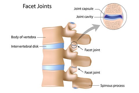 A joint is a connection between two bones. Our spine is made up of multiple joints at each level and many of these joints in the spine are synovial joints. Synovial joints are the most common and most movable type of joint in our body. Synovial joints have a joint capsule surrounding the bony surfaces of the joint and lubricating synovial fluid in the joint capsule. The skull is connected to the cervical spine at C1 by synovial joints called the occipital-cervical joint. Each spinal motion segment of the spine has a pair of facet joints that provide the posterior support for the spine. A pivot joint is found at the very top of the spine, called the atlanto-axial joint. This joint is between the very first and second cervical vertebrae which are very specialized to the spine to allow for rotational movement.
 -
MOTION COMPLEX
Each level of your spine functions as a three-joint complex. There are two facet joints in the back and a large disc in front. This tripod creates great stability, supports all your weight above each level and provides support for you to move in all directions. Vertebrae are stacked one on top of another and are separated by intervertebral discs, which act as an elastic cushions or shock absorbers. The first two cervical vertebrae are an exception and do not have discs. The interbody space is the disc space that is located between the vertebral body bones.
-
CERVICAL VERTEBRAE
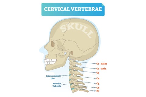 There are seven cervical vertebrae, named C1-C7, designed for flexibility and movement. The cervical spine has a lordotic shape, or a backwards “C” shape. The first two cervical vertebrae are very specialized to allow us to turn our head from side to side. The first cervical vertebra (C1) is called the atlas and is aptly named after the Greek god Altas, who carried the world on his shoulders. This bone is formed like a ring that sits upon the second cervical vertebra (C2). The second cervical vertebra (C2) is called the axis as it is the line upon which the head and C1 rotate upon. The C1 vertebra connects the skull to the cervical spine. These two vertebrae have different anatomy than the rest of the spine. The C1 vertebra is formed like a ring that sits on top of C2. The C2 vertebra has a bony knob that fits into the front portion of the ring of the C1 vertebra. This bony knob is called the odontoid process or dens. It is held in place by a special ligament that holds it tightly to the front of the ring of the C1 vertebra. While the cervical spine is very flexible, it is also at greatest risk for injury from strong sudden movements, as there is less muscular support.
 -
THORACIC VERTEBRAE
There are twelve thoracic vertebrae, named T1-T12, specialized for stability. The thoracic spine aids in keeping the body upright, protects vital chest organs and articulates with each rib to form the rib cage. Each rib is firmly connected to each level of the thoracic spine. The thoracic spine has a kyphotic shape, a “C” shape, and the discs in this part of the spine are relatively thin.
-
LUMBAR VERTEBRAE
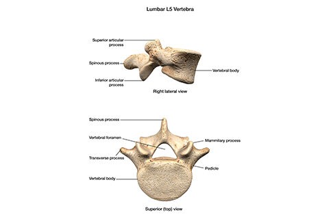 There are usually five lumbar vertebrae, named L1-L5, designed for weight bearing loads and movement. In some people, they may have developed four or six lumbar vertebrae. In some cases one of the bones of the sacrum, the base of the spine, forms as a vertebra instead of the sacrum. This is called a transitional (or sixth) vertebra and is simply a bony anomaly.
The lumbar spine is shaped like the cervical spine; it is lordotic like a backwards “C”. The two lordotic curves in the neck and low back are balanced by the thoracic kyphotic “C” curve so the spine’s center of gravity is overall balanced in an “S” shape. The vertebrae in the lumbar spine are the largest of the entire spine, designed to hold increasing forces of weight. The lumbar spinal canal is also the largest, allowing for more space for the nerves.
-
SACRUM AND COCCYX
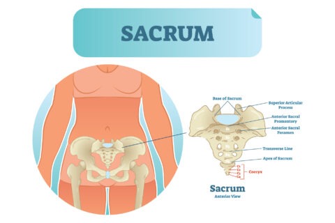 The sacrum is made of five fused vertebrae that form a single bone. The sacrum is shaped like an inverted triangle with the base at the top. It acts as a wedge between the two iliac pelvic bones. On both sides of the pelvis, the sacrum articulates with the ilium through the sacroiliac joints. The coccyx is formed by the fusion of four to five rudimentary vertebrae, commonly referred to as the tailbone.
 -
SACROILIAC JOINT
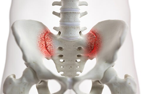 The sacroiliac (SI) joint is a strong weight bearing joint in the pelvis that connects the sacrum and pelvis. There are two joints, one on each side of the sacrum. This joint is reinforced by strong surrounding ligaments as shown in the image. Both joints move together as single unit to transmit upper body forces and provide shock absorption for the spine. A series of ridges and valleys in the joint fit together like a lock and key, much like if you put your knuckles together. There is a small amount of movement in this joint to allow for a walking gait pattern in normal human locomotion. Just like other joints in the body, this joint can become inflamed, unstable and dysfunctional.
 -
INTERVERTEBRAL DISC
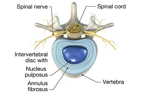 An intervertebral disc is a strong ligament that connects one vertebral bone to the next. The discs are the shock-absorbing cushions between each vertebra of the spine. The disc is made up of three basic structures: the annulus fibrosus, the nucleus pulposus, and the vertebral end-plates. All three disc structures are made of different percent compositions of proteoglycan (proteins that bind water), collagen (the main protein in connective tissue) and water. The varying compositions create different functions. All these properties make the disc unique and highly specialized. Discs in the spine increase in size from the neck to the low back as there are increasing needs for shock absorption due to weight and gravity. These specific disc ligaments function just like knee ligaments and shoulder ligaments do. They allow the spine to move so we can bend forward, backward and sideways. Just like other ligaments, the discs can be injured. The intervertebral disc is essential for providing spinal stability and proper alignment, withstanding compressive shock forces, and allowing for movement between vertebral bones. The intervertebral disc is an avascular tissue, meaning it does not naturally have a blood supply. Capillaries from the surface of the vertebral bodies supply the disc with nutrients. Thus, the disc is dependent on the blood supply to the vertebral bones. Each disc has a strong outer ring of fibers, called the annulus fibrosus. The annulus is comprised of specialized sheets of connective tissue, called lamellae, which are layered for strength. The annulus has a higher amount of collagen that makes the outer layer extremely stiff. The nucleus pulposus is the soft, jelly-like center. It serves as the main shock absorber and is held in place by the outer annulus. The nucleus consists of a gel like matrix that provides maximum hydration. It functions to distribute pressures in all directions within each disc under compressive forces.
 -
SPINAL CORD AND NERVES
The spinal cord connects the nervous system from the brain to the rest of the body. The spinal cord leaves the brain through a hole in the base of the skull called the foramen magnum. The spinal cord and nerves travel from the cervical spine down to the lowest point of the spine, the sacrum. Spinal nerves exit the spinal canal between the vertebrae at each level. Two nerves exit each level, one on the left and one on the right. These nerves exit through openings called foramen. The nerves leave the spinal cord and travel to specific destinations in the body. The nerves leaving the neck travel to both arms and those from the low back to each leg. The specific pattern each nerve innervates is called a dermatome pattern.
Meet our Doctors

Dr. Santoshi Kurada
MBBS, MD, Fellow in Critical Care and Pain Medicine (UK)

Dr. Swagatesh Bastia
MBBS, MS (Ortho), Fellow in Trauma and Orthopaedics (UK), Fellow in Pain Management

Dr. Faraz Ahmed Syed
MBBS, MS (Orthopaedics), AO Spine Surgery Fellow (UK)

Dr. Wiquar Ahmed
MBBS, DA, FIPM, CIPS

Dr Roshan Adappa
MBBS, MD (Anaesthesiology), FIASP, MMed -Pain Management (UK), European Diploma in Pain Medicine (EDPM, Belgium)

Dr. Shubha V Hegde
MBBS, MD, DNB(Anaesthesia), EDPM-1 (Europe), FIAPM, FIPM, CCEPC
