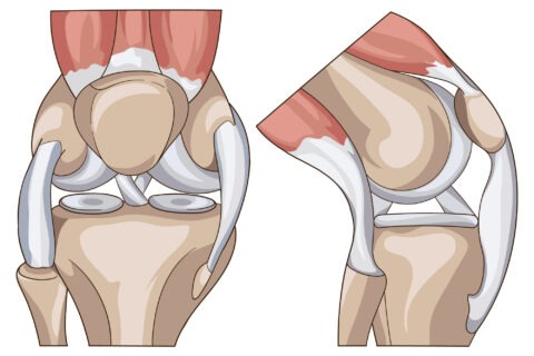
The knee joint is a hinge type synovial joint, which mainly allows for flexion and extension (and a small degree of medial and lateral rotation). It is formed by articulations between the patella, femur and tibia.
Anatomy of the Knee
Anatomy of the Knee
Anatomy of the Knee
Knee is the largest hinge joint of the body and is lined by an articular cartilage which is lubricated by synovial fluid. It is formed by the articulation of the distal/lower end of the femur(thigh bone) and the the proximal/upper end of the tibia(shin bone).
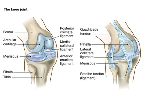
Quadriceps tendon connects the quadriceps muscle to patella(knee cap). Patellar tendon connects the patella onto the tibia.
Ligaments join the bones forming the knee joint and provide stability to the knee:
* The anterior cruciate ligament prevents the femur from sliding backward on the tibia (or the tibia sliding forward on the femur).
* The posterior cruciate ligament prevents the femur from sliding forward on the tibia (or the tibia from sliding backward on the femur).
* The medial and lateral collateral ligaments prevent the femur from sliding side to side.
Two C-shaped pieces of cartilage called the medial and lateral menisci act as shock absorbers between the femur and tibia.
Knee is surrounded by numerous bursae, which are fluid filled spaces and help in smooth movement of the knee.
Meet our Doctors

Dr. Santoshi Kurada
MBBS, MD, Fellow in Critical Care and Pain Medicine (UK)

Dr. Swagatesh Bastia
MBBS, MS (Ortho), Fellow in Trauma and Orthopaedics (UK), Fellow in Pain Management

Dr. Faraz Ahmed Syed
MBBS, MS (Orthopaedics), AO Spine Surgery Fellow (UK)

Dr. Wiquar Ahmed
MBBS, DA, FIPM, CIPS

Dr Roshan Adappa
MBBS, MD (Anaesthesiology), FIASP, MMed -Pain Management (UK), European Diploma in Pain Medicine (EDPM, Belgium)

Dr. Shubha V Hegde
MBBS, MD, DNB(Anaesthesia), EDPM-1 (Europe), FIAPM, FIPM, CCEPC

Dr. Pramodh
MBBS,DFM,CCEBDM,MLSM
© 2020-21 Alleviatepainclinic. All rights reserved.