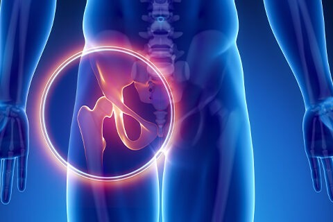
anatomy hip 1

Anatomy of Hip

Anatomy of Hip

The Hip Joint The hip joint is a ball and socket type joint.

The femoral head (ball) fits into the acetabulum (socket) of the pelvis. The large round head of the femur rotates and glides within the acetabulum. The depth of the acetabulum is further increased by a fibrocartilagenous labrum that attaches to the outer rim of the acetabulum. It acts to deepen the socket and to add additional stability to the hip joint.  The labrum can become torn and cause symptoms such as pain, weakness, clicking, and instability of the hip.

anatomy hip 2


  • Bones of the Hip Joint

    • There are numerous structures that contribute stability to the hip:
    • The ball and socket bony structure
    • The labrum
    • The capsule and its associated ligaments: e.g. iliofemoral ligament, pubofemoral ligament
    • The surrounding muscles including the abductors (gluteus medius and minimus) and external rotators (gemelli muscles, piriformis, the obturators).
  • Hip Ligaments

    The iliofemoral ligament in the hip
    The stability of the hip is increased by the strong ligaments that encircle the hip (the iliofemoral, pubofemoral, and ischiofemoral ligaments). These ligaments completely encompass the hip joint and form the joint capsule. The iliofemoral ligament is considered by most experts to be the strongest ligament in the body. The ligamentum teres is a small tubular structure that connects the head of the femur to the acetabulum. It contains the artery of the ligamentum teres. In infants, this serves as a relatively important source of blood supply to the head of the femur. In adults, the ligamentum teres is thought by most to be more of a vestigial structure that serves little function.

  • The ischiofemoral ligament of the hip

    Muscles of the Hip
    The muscles of the thigh and lower back work together to keep the hip stable, aligned and moving. It is the muscles of the hip that allow the movements of the hip:
    flexion – bend
    extension – straighten
    abduction – leg move away from midline
    adduction – leg moves back towards midline
    external rotation (allows for the foot to point outwards)
    internal rotation (allows for the foot to point inward)
    The hip muscles are divided up into three basic groups based on their location: anterior muscles (front), posterior (back), and medial (inside). The muscles of the anterior thigh consist of the quadriceps (or quads): vastus medialis, intermedius, lateralis and rectus femoris muscles. The quads make up about 70% of the thigh’s muscle mass. The main functions of the quads are flexion (bending) of the hip and extension (straightening) of the knee.
    The gluteal and hamstring muscles, as well as the external rotators of the hip are located in the buttocks and posterior thigh. The gluteal muscles consist of the gluteus maximum, gluteus medius, and gluteus minimus. The gluteus maximus is the main hip extensor and helps keep up the normal tone of the fascia lata or iliotibial (IT) band, which is the long, sheet-like tendon on the side of your thigh. It helps with motion of the hip, but perhaps more importantly, acts to help stabilize the knee joint.
    Gluteus medius and minimus are the main abductors of the hip —that is, they move the leg away from the midline of the body (using the spine as a midline reference point). They also are the main internal rotators of the hip (i.e. turn the foot inwards). The gluteus medius and minimus are also important stabilizers of the hip joint and help to keep the pelvis level as we walk.
    The tensor fascia lata (TFL) is another abductor of the hip, which, along with the gluteus maximus, attaches to the IT band. The IT band is a common cause of lateral (outside) hip, thigh, and knee pain.
    The medial muscles of the hip are involved in the adduction of the leg i.e. bringing the leg back towards the midline. These muscles include the adductors (adductor magnus, adductor longus, adductor brevis, pectineus, gracilis). Obturator externus also helps to adduct the leg.
    The external rotator muscles (piriformis, gemelli, obturator internus) of the hip are located in the buttock area and assist in lateral rotation of the hip (out-toeing). Lateral rotation is needed for crossing the legs.

  • Blood Vessels and Nerves of the Hip

    The sciatic nerve is located where it could get injured from a backwards dislocation of the femoral head.
    Nerves carry signals from the brain to the muscles to move the hip and carry signals from the muscles back to the brain about pain, pressure and temperature. The main nerves of the hip that supply the muscles in the hip include the femoral, obturator, and sciatic nerves.
    The sciatic nerve is the most commonly recognized nerve in the hip and thigh. The sciatic nerve is large—as big around as your thumb—and travels beneath the gluteus maximus down the back of the thigh where it branches to supply the muscles of the leg and foot. Hip dislocations can cause injury to the sciatic nerve.
    The blood supply to the hip is extensive and comes from branches of the internal and external iliac arteries: the femoral, obturator, superior and inferior gluteal arteries. The femoral artery is well-known because of its use in cardiac catheterization. You can feel its pulse in your groin area. It travels from deep within the hip down the thigh and down to the knee. It is the continuation of the external iliac artery which lies within the pelvis. The main blood supply to the femoral head comes from vessels that branch off of the femoral artery: the lateral and medial femoral circumflex arteries. Disruption of these arteries can lead to osteonecrosis (bone death) of the femoral head. These arteries can become disrupted with hip fractures and hip dislocations.

  • Bursae

    Bursae are fluid filled sacs lined with a synovial membrane which produce synovial fluid. Bursae are often found near joints. Their function is to lessen the friction between tendon and bone, ligament and bone, tendons and ligaments, and between muscles. There are as many as 20 bursae around the hip. Inflammation or infection of the bursa called bursitis.
    The trochanteric bursa is located between the greater trochanter (the bony prominence on the femur) and the muscles and tendons that cross over the greater trochanter. This bursa can get irritated if the IT band is too tight. This bursa is a common cause of lateral thigh (hip) pain. Two other bursa that can get inflamed are the iliopsoas bursa, located under the iliopsoas muscle and the bursa located over the ischial tuberosity (the bone you sit on).

  • Common Problems of the Hip

    • Posterior Hip dislocation
    • Osteoarthritis
    • Dislocation (see image above of simple dislocation)
    • Bursitis
    • Fracture
    • Femoroacetabular impingement
    • Labral tear
    • IT band syndrome
    • Snapping hip syndrome
    • Aseptic or Avascular necrosis
    • Congenital Dislocation
    • Acetabular dysplasia
    • Coxa valga
    • Coxa vara
      Tumor
      Legg-Perthes disease

Meet our Doctors